Published:
An American physicist has won the Nobel Prize for his pioneering work developing an optical microscopy technique that is now behind an exciting research project involving Heriot-Watt University.
Arthur Ashkin shares the 9m Swedish Kronor prize alongside Canadian scientist, Donna Strickland and Gerard Mourou from France who, this week, were named as the 2018 Nobel Prize winners in Physics.
Ashkin won the coveted title for his groundbreaking work in developing 'optical tweezers' that allow scientists to capture and move cells, bacteria and even viruses using the power of light alone.
Optical tweezers are at the centre of a new £1m, three-year project, between the University of Nottingham, Heriot-Watt University and the University of Glasgow to develop a state-of-the-art optical instrument to better understand the spread of cancer and human disease.
If successful, the instrument will give scientists a unique 4D-view of live cells, allowing them greater insight into the triggers that lead to mutation and why disease spreads around the human body.
Dr. Amanda Wright, Project Lead from the University of Nottingham, said: “This instrument will revolutionise our understanding of how cells respond to the forces imposed on them by their matrix as they move through it - forces which directly control cell function and behaviour.
"For the first time, we will be able to view the cellular microworld from a cell's own perspective. It will help us to learn how migrating cancer cells, for example, exploit existing tracts and directional cues inside the matrix."
The research will result in a bespoke instrument, combining four cutting-edge optical microscopy technologies in a way that has not been previously possible, creating a single multifunctional platform.
The tool will track and analyse the cells' position, movement and function as they grow into and interact with the proteins and sugars that make up their surrounding environment (the extracellular matrix).
This will enable scientists to understand how physical and biochemical cues from cells influence the matrix, and vice versa, and how these interactions affect the progression of disease.
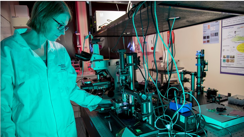
Dr Paul Dalgarno from Heriot-Watt University who pioneered multiplane imaging technique that for three dimensional imaging, said: “While a conventional microscope produces a sharp image from just a single object-plane, multiplaning records sharp images from several object-planes simultaneously.
“We will combine our revolutionary multiplane system with optical trapping before integrating this into the full light sheet based trapping system of our co-collaborators.
Here at Heriot-Watt, we already have an established record for optical trapping and this project aims to take that expertise and use it as the basis to help probe 3D cell growth. Together these techniques will enable us to fully study how cells migrate, grow and interact in 3D and in real time.”
The new approach will be important for developing therapies for diseases where cells respond abnormally to signals from their local matrix, such as cancer. The advancement in optical technology could also benefit research into degenerative diseases, regenerative medicine and infection pathways as scientists build a better understanding that will ultimately inform treatments.
A second research strand will examine how cells absorb particles and the impact that drug therapies (such as functionalised polymer nanoparticles) have on the matrix. The findings could help to design new medicines and drug delivery systems targeted at the macromolecular level.
Commenting on the collaboration, Professor John Underhill, Chief Scientist at Heriot-Watt University, said: “This million pound investment in life sciences research is a demonstration of the potential and importance of collaboration in research in general and this exciting research theme in particular.
"At Heriot-Watt University, we pride ourselves on being an international university with a global outlook, finding solutions to challenges with wide ranging applications. Our cutting-edge research into microscopy could have wide reaching implications for the treatment of some of the world's biggest killer diseases including cancer.”
The research project is being funded by The Engineering and Physical Sciences Research Council (EPSRC) with co-funding from the Medical Research Council (MRC) and the Biotechnology and Biological Sciences Research Council (BBSRC).